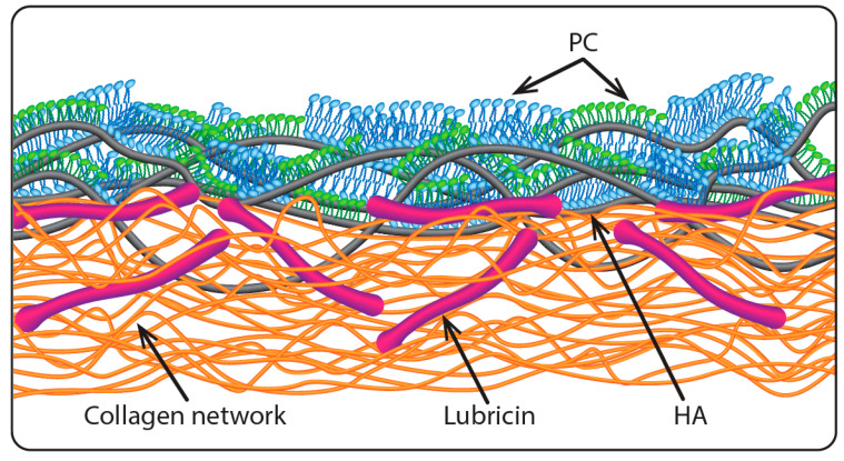
Figure 1.
Illustrating the proposed boundary layer on articular cartilage [33], where lubricin molecules anchor hyaluronic acid (HA) chains at the articular cartilage surface and the HA is complexed with phosphatidylcholine (PC) layers exposing their highly hydrated phosphocholine groups at the very outer surface (the slip plane). Reproduced with permission from [33]. Copyright 2016, Annual Review.

