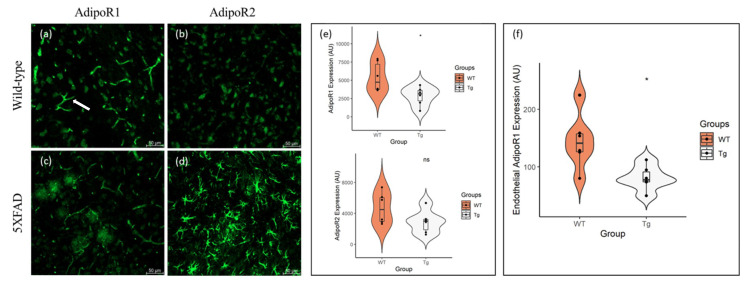
Figure 1.
Expression of AdipoR1 and AdipoR2 in the cortices of 48–52-week-old 5XFAD and wild-type (WT) mice. AdipoR1 is present in neurons of both (a) WT and (c) 5XFAD mice, in addition to endothelial cells lining blood vessels (arrow). Both (b) WT and (d) 5XFAD mice also express AdipoR2 in neurons, with substantial glial expression of AdipoR2 in (d) 5XFAD mice. There was a reduction in (e) neuronal AdipoR1 and AdipoR2 expression in the 5XFAD (Tg) cortex, compared to controls, with statistical significance observed in AdipoR1 expression. Endothelial expression (f) of AdipoR1 was also significantly reduced in the 5XFAD (Tg) cortex. Data are presented as Mean ± SEM using independent t-test with statistical significance * p < 0.05 denoted between groups. Images were taken at 20× magnification. Scale bar = 50 μm.

