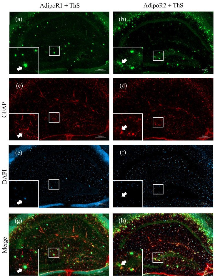
Figure 4.
Amyloid plaque staining and adiponectin receptor expression in the hippocampi of 48–52-week-old 5XFAD mice. AdipoR1 (a) double-labelled with GFAP (c) and counterstained with 4′,6-diamidino-2-phenylindole (DAPI) (e) displayed no AdipoR1-expressing astrocytes surrounding Thioflavin-S-stained amyloid plaques (arrows) in the hippocampus (g). Astrocytes expressing AdipoR2 (b) double-labelled with GFAP (d) and counterstained with DAPI (f) can be seen surrounding amyloid plaques (arrows) (h). Images were acquired at 20× magnification. Scale bars for (a–h) are 200 μm. Scale bars for zoomed-in figures are 20 μm.

