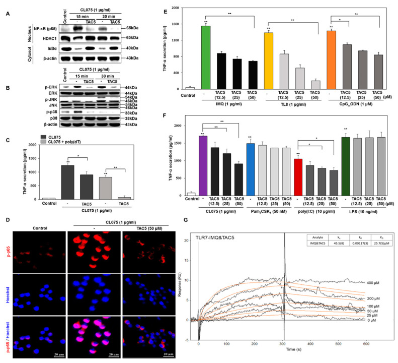
Figure 2.
TAC5 inhibits the CL075-induced proinflammatory response. RAW 264.7 cells were treated with 50 µM of TAC5 for 1 h, followed by treatment with CL075 (1 µg/mL) for 15 and 30 min, respectively. (A) Nuclear factor kappa-light-chain-enhancer of activated B cells (NF-κB) activation (NF-κB translocation into the nucleus and degradation of IκBα in the cytosol) was measured by western blotting. Histone deacetylase-1 was used as a nucleus loading control. β-actin was used as a cytosol loading control. (B) The expression levels of mitogen-activated protein kinases (MAPKs) were evaluated by western blot analysis using whole protein extracts, with inactive MAPKs used as controls (β-actin was used as a loading control). (C) Inhibition of TNF-α secretion by TAC5 in the presence of CL075 and a combination of CL075 and poly(dT). (D) Confocal microscopy image of the phosphorylated NF-κB (p-p65) expression level. The red staining corresponds to phosphorylated p65 subunit of NF-κB (p-p65) and blue staining (Hoechst33258) indicates nuclei (the scale bar is 20 µm in width). (E,F) The expression level of TNF-α was measured by ELISA when the TAC5 was co-treated with (E) imiquimod (TLR7), TL8-506 (TLR8), CpG ODN (TLR9) (F) Pam3CSK4 (TLR1/2), poly(I:C) (TLR3), LPS (TLR4), or CL075 (TLR7/8) agonists in RAW 264.7 cells. (G) Surface plasmon resonance (SPR) sensorgram illustrating the competitive binding of TAC5 to TLR7 in the presence of imiquimod. The data shown represent five independent experiments, and the bars represent means ± SEM (* p < 0.05, ** p < 0.01).

