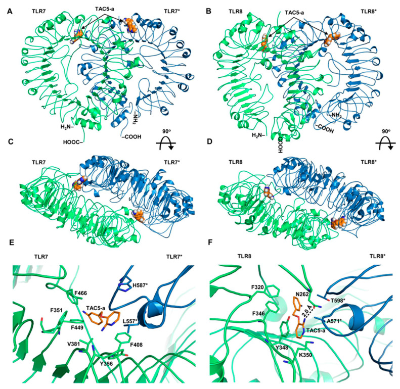
Figure 7.
Structural models of TAC5-a bound to TLR7 and TLR8. (A) The overall structure of a homology model of human TLR7 having TAC5-a at the synthetic agonist binding cavity. (B) The structure of human TLR8 with TAC5-a bound to the agonist-binding site. (C) The top surface of TLR7 after 90° rotation of panel (A). (D) A 90°-rotated view of the panel (B). (E) Detailed intermolecular interaction of TAC5-a with the amino acids of TLR7. ‘*’ indicates chain B of the protein. (F) A close-up view of the intermolecular contacts between TAC5-a and TLR8 residues. ‘*’ indicates subunit B of the protein. The hydrogen bond (H-bond) is shown as a dashed line. The digit indicates the distance of the H-bond in Å. TAC5-a is shown as an orange stick. The chain As of both proteins are colored lime green and the chain Bs are sky blue.

