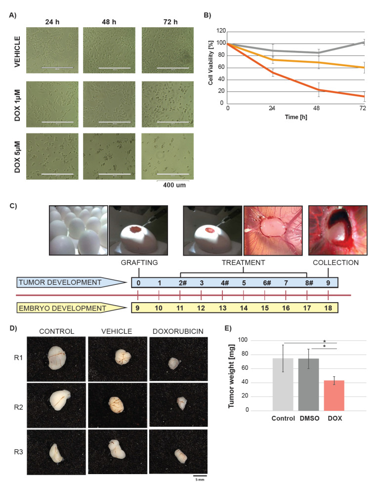
Figure 1.
Doxorubicin decreases cell viability in ovo and in vitro. (A) Representative appearance of the MB-MDA-231 cultured in vitro over time of 72 h after treatment with vehicle (DMSO) and two doxorubicin (DOX) concentrations. (B) Cell viability after the DMSO or DOX treatment depicted by the (3-(4,5-dimethylthiazol-2-yl)-2,5-diphenyltetrazolium bromide) MTT assay. Dark grey indicates vehicle, yellow and red indicate 1 µM and 5 µM concentration of DOX respectively. (C) Study design. The chick embryo grew over a period of nine days. At day 9 of embryo growth, the cancer cells from the MB-MDA-231 cell line were grafted on the CAM of the egg. The treatment with DMSO, and DOX started at day 2 after cancer cell grafting. A volume of 100 µL of 25 µM DOX was added to achieve 0.024 mg/km of DOX per egg. The untreated cells were used as a control. The treatment was conducted every two days and the treatment time points are depicted by #. The tumors were collected at day 9 after the cancer cell grafting. (D) Representative gross appearance of tumors excised from the chick embryo (each group presented in triplicates). (E) Impact of DOX on tumor weight. Light grey indicates control, dark grey indicates vehicle, and red indicates DOX treatment. Significant differences were depicted by *.

