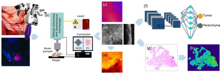
Figure 6.
Workflow for the processing of CNS tumor biopsies. The samples were resected during 5-ALA FGS [13] (a) and immediately transferred to the lab, where FI-OCM imaging was performed (b). FI data were acquired before bleaching occurred (c), followed by OCM imaging (d). Based on OCM intensity data, attenuation maps were computed (e). Data post-processing was performed and all OCM data sets were evaluated using a convolutional neural classifier model (f). Subsequently, the samples were fixed in 4% formalin and processed for histopathological workup. H&E staining (g) and cell density maps (h) served as ground truth.

