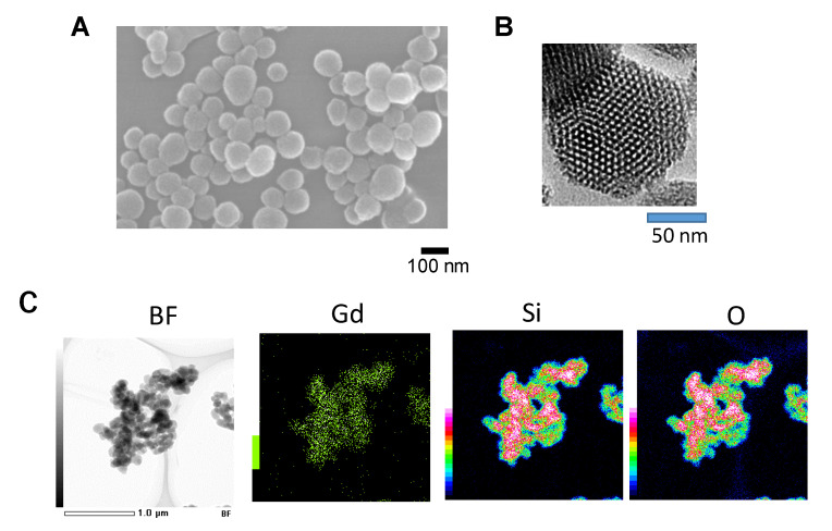
Figure 2.
Mesoporous silica nanoparticles (MSN) prepared by the sol–gel method. (A) Scanning electron microscope (SEM) picture; (B) Transmission electron microscope (TEM) picture; (C) Scanning transmission electron microscopy-energy dispersive X-ray (STEM-EDX) images of Gd–MSN. Bright field image as well as elemental mapping images of Gd, Si and O are shown. Modified from [9].

