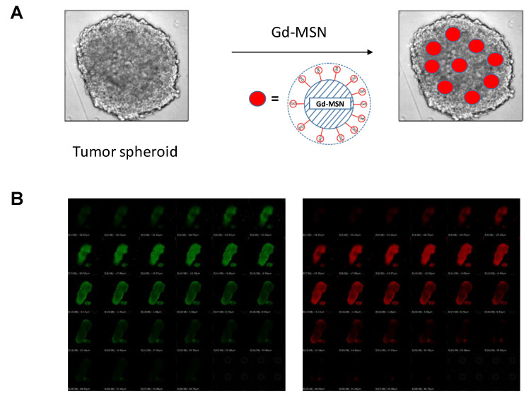
Figure 3.
(A) Schematic drawing showing tumor spheroids incubated with Gd–MSN results in the distribution of Gd–MSN throughout the spheroid. Position of Gd–MSN in the spheroid is arbitrary to emphasize the presence of Gd–MSN in the spheroid; (B) tumor spheroid prepared from GFP-expressing cancer cells is observed as a green mass. When the spheroid was incubated with gadolinium-loaded MSN labeled with rhodamine B, uniform distribution of the nanoparticle throughout the spheroid was observed by using confocal microscopy (see the red fluorescence). Focal planes from the top to the bottom of the spheroid sample is shown. In all planes, green fluorescence of the cancer cells overlaps with the red fluorescence of Gd–MSN. Modified from [9].

