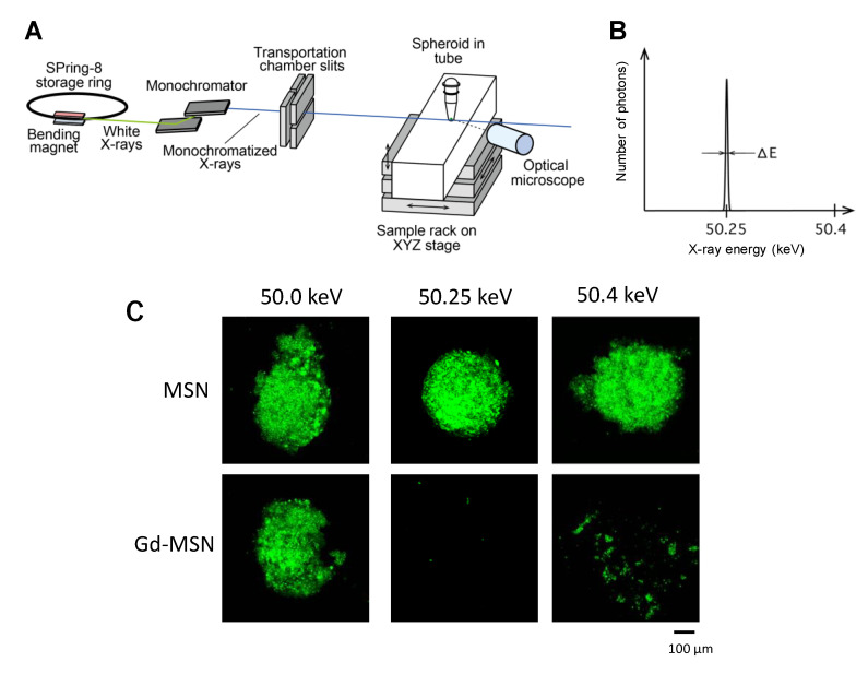
Figure 4.
Irradiation with monochromatic X-ray. (A) Experimental setup; (B) band width of the 50.25-keV monochromatic X-ray; (C) tumor spheroids incubated with Gd–MSN were irradiated with 50.0, 50.25- or 50.4-keV monochromatic X-ray for 20 min and then incubated for three days. Tumor spheroids with Gd–MSN after irradiation with 50.25 or 50.4 keV were destructed, while spheroids with empty MSN were not affected by the irradiation. Modified from [9]

