Figure 2.
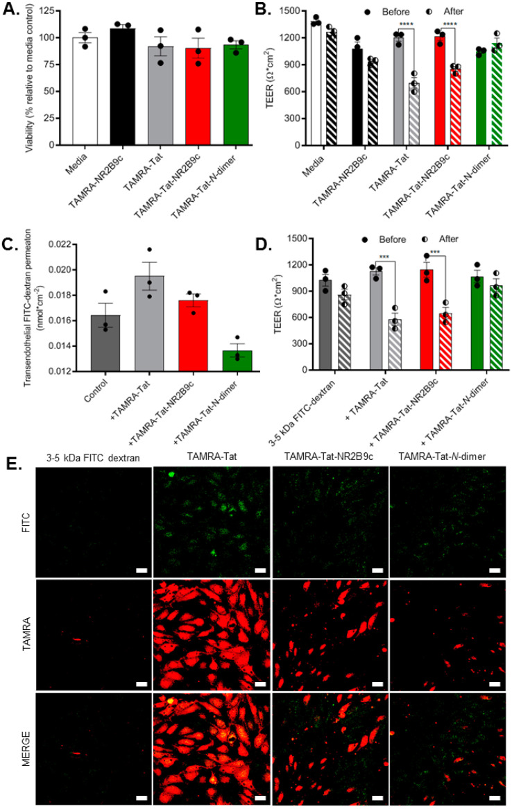
(A) The cellular viability was assessed following application of 100 µM TAMRA-NR2B9c, TAMRA-Tat, TAMRA-Tat-NR2B9c, or TAMRA-Tat-N-dimer to the in vitro BBB model for 3 h. (B) The barrier integrity was evaluated by measuring transendothelial electrical resistance (TEER) before and after application of the peptides (100 µM). (C) Transport of 3–5 kDa FITC-dextran was quantified after 3 h of incubation on the in vitro BBB model alone (1 mg/mL; control) or in the presence of the peptide (100 µM). (D) The barrier integrity was assessed by measuring TEER before and after application of 4 kDa FITC-labelled dextran (1 mg/mL) together with the peptide (100 µM). Data are presented as mean ± SEM (N = 3, n = 3). *** p < 0.001 and **** p < 0.0001 (one-way ANOVA with Dunnett’s multiple comparisons test). (E) Representative confocal microscopy images of 4 kDa FITC-dextran uptake into the endothelial cells after 3 h of incubation and cell fixation. Maximal z-stack projections, scale bar: 20 µm.

