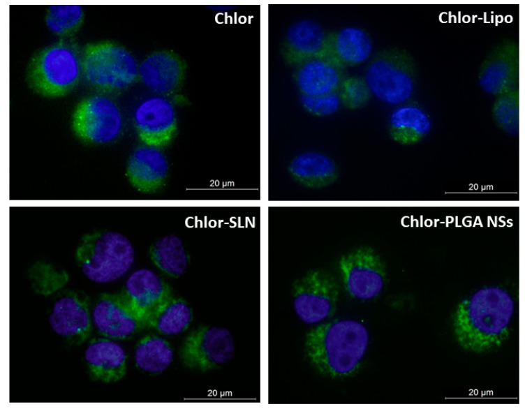
Figure 4.
Representative fluorescence images of PC-3 cells incubated with the various Chlor formulations. Cells were incubated for 6 h with Chlor-Lipo or Chlor-SLN and for 24 h with Chlor or Chlor-PLGA NSs at the same Chlor concentration (5 μM); DAPI (blue) was used a nuclear counterstain. Magnification: 63×. Scale bars: 20 μm.

