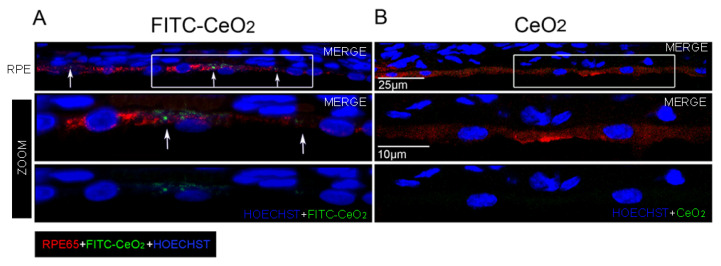
Figure 2.
Localization of cerium oxide nanoparticles in the retinal pigment epithelium. Representative confocal images of retinal cryosections of albino rats immunolabeled with anti-RPE65 (red) in order to detect the retinal pigment epithelium: (A) intravitreally injected with fluorescein-isothiocyanate (FITC-CeO2) (green), the white arrows indicate the FITC-CeO2 agglomerates, which localize in the retinal pigment epithelium (RPE); (B) Intravitreally injected with standard cerium oxide nanoparticles. The high magnifications show the regions highlighted in the white frames. CeO2: cerium oxide nanoparticles. FITC-CeO2: cerium oxide nanoparticles labeled with FITC.

