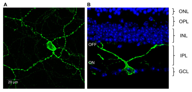
Figure 6.
Visualization of melanopsin expressing photosensitive retinal ganglion cells (pRGCs) of the adult mouse retina. (A) Confocal microscopy image of a flat mounted mouse retina showing antibody labeling of melanopsin in an M1 type pRGC with high levels of melanopsin expression and large but sparse dendritic fields. Additionally, note the presence of a weaker stained processes from neighboring non-M1 cells. (B) Cross section image of the mouse retina showing the dendrites of M1 type pRGCs extending to the OFF layers of the inner plexiform layers (IPL). Abbreviations: Outer nuclear layer (ONL); inner nuclear layer (INL); inner plexiform layer (IPL); ON and OFF mark the ON and OFF sublaminae of the IPL; ganglion cell layer (GCL).

