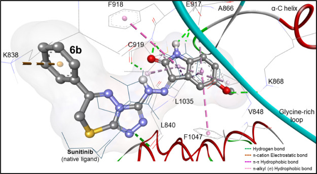
Figure 6.
Docking mode of compound 6b (in ball and stick view) into the binding site of VEGFR-2 (PDB: 4agd). It seems to be superimposed on the native ligand (B49; sunitinib in dark turquoise lines) within a root-mean-square deviation (rmsd) of 1.14 Å, revealing hydrogen, electrostatic, and hydrophobic binding interactions identical to sunitinib.

