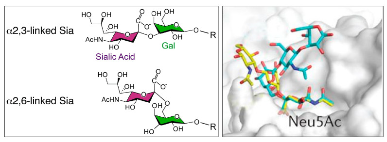
Figure 1.
(Left): Structure of N-acetylneuraminic acid (Neu5Ac, sialic acid, SA), linked to galactose either via a α-2,3- or via a α-2,6-linkage. Figure taken from [18], which is an open access article distributed under Creative Commons attribution license. (Right): Superposition of an avian influenza virus hemagglutinin in a complex with α-2,3-sialyllactosamine (yellow) (PDB accession 2WR2) and the human receptor α-2,6-sialyllactosamine (cyan) (PDB accession 2WR7). The avian receptor generally has a linear conformation, whereas the human receptor is more flexible and has an umbrella-like topology. Reproduced with permission from [14]. Copyright Nature, 2014.

