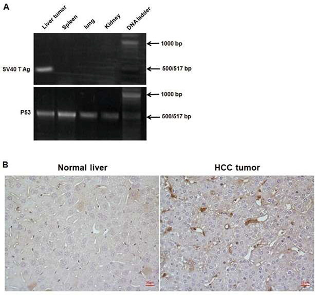
Figure 5: Specific detection of SV40 T antigen gene in tumor.
(A) Tumor tissue and additional tissue samples throughout mice were collected from tumor-established mice. Genomic DNA was isolated from tissue and primers for both SV40 T Ag and P53 (control) were synthesized for conventional PCR. SV40 T antigen gene was detected in tumor tissue but is not present in either the normal tissue or other tissue in the mice. (B) Tumor tissue was collected from tumor-bearing mice and stained with anti-SV40 T antigen antibody. Left panel is a negative control from healthy liver tissue collected from naïve mice. Right panel shows significant SV40 T antigen staining of tumor tissue collected from tumor-bearing mice (brown color). Magnification = 40x objective, scale bar = 20 μm. Please click here to view a larger version of this figure.

