
Figure 2.
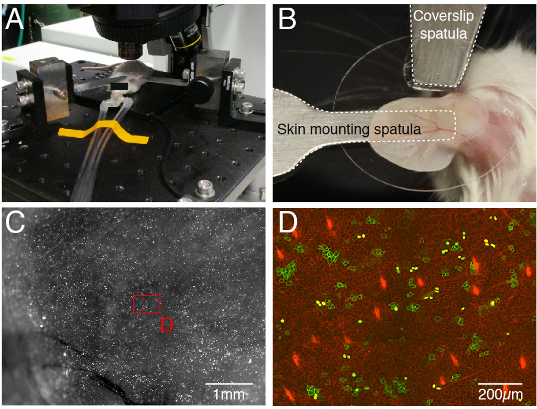
Mouse mounting. An anesthetized mouse on stage of confocal microscope. Anesthetic is delivered through nose cone (A). For ear mounting two spatulas are used, one to support the tissue form below (skin mounting spatula) and a second one to which the coverslip is glued placed on top (coverslip spatula). The ear is held firmly in place between the both spatulas (B). Ear after clonal induction at high magnification, shown is green channel (C). High magnification image of ear epidermis after clonal induction. Membrane EGFP (“normal clones”) and nuclear EGFP (“Gof/Lof clones”) are easily identifiable among red membrane-labeled cells (D).