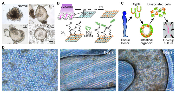
Figure 3.
The microfluidic culture of the patient-derived organoid epithelium in a PMI Chip. (A) The phase contrast micrographs of human intestinal organoids derived from a normal donor or the patients diagnosed with UC, CD, and CRC. (B) The experimental procedure for the activation of a microfluidic channel and a subsequent ECM coating in a PMI Chip. (C) The procedure of the formation of biopsy-derived intestinal organoids and its use for a microfluidic culture in a PMI Chip. (D) The phase contrast images that show the 3D regeneration of patient-derived intestinal organoid epithelium under non-linear luminal flow and multiaxial stretching motions. Normal organoid cells were cultured in a PMI Chip for 7 days under flow (50 µL/h) and stretching motions (5%, 0.15 Hz). Bars, 200 μm.

