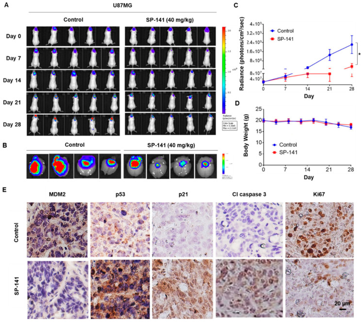
Figure 3.
SP-141 shows excellent antitumor activity against U87MG human glioblastoma intracranial xenografts. (A) Representative images of bioluminescence acquisition of the brain tumors in nude mice after 0, 7, 14, 21, and 28 days of treatment. SP-141 was administered intraperitoneally at 40 mg/kg/day, 5 days per week for 4 weeks. SP-141 was dissolved in PEG400/EtOH/saline (57.1:14.3:28.6, v/v/v). The control group received the vehicle alone. (B) Ex-vivo bioluminescence from the brains of control (left panel) and SP-141-treated (right panel) mice. The scale indicates the fluorescence intensity in terms of radiant efficiency (p/sec/cm2/Sr). (C) Average time-dependent changes in bioluminescence radiance in U87MG xenografts in mice receiving SP-141 therapy over 4 weeks (* p < 0.01). (D) The average body weights of mice were plotted during SP-141 treatment. (E) Reduced MDM2 expression and activation of p53 signaling components were observed in U87MG intracranial xenografts following SP-141 treatment. Brain areas bearing the U87MG tumors from control and SP-141 treated nude mice were sectioned and processed for the expression of MDM2, p53, p21, cleaved caspase 3, and Ki67 proteins by immunohistochemistry. Representative staining patterns are shown. Scale bars: 20 µm.

