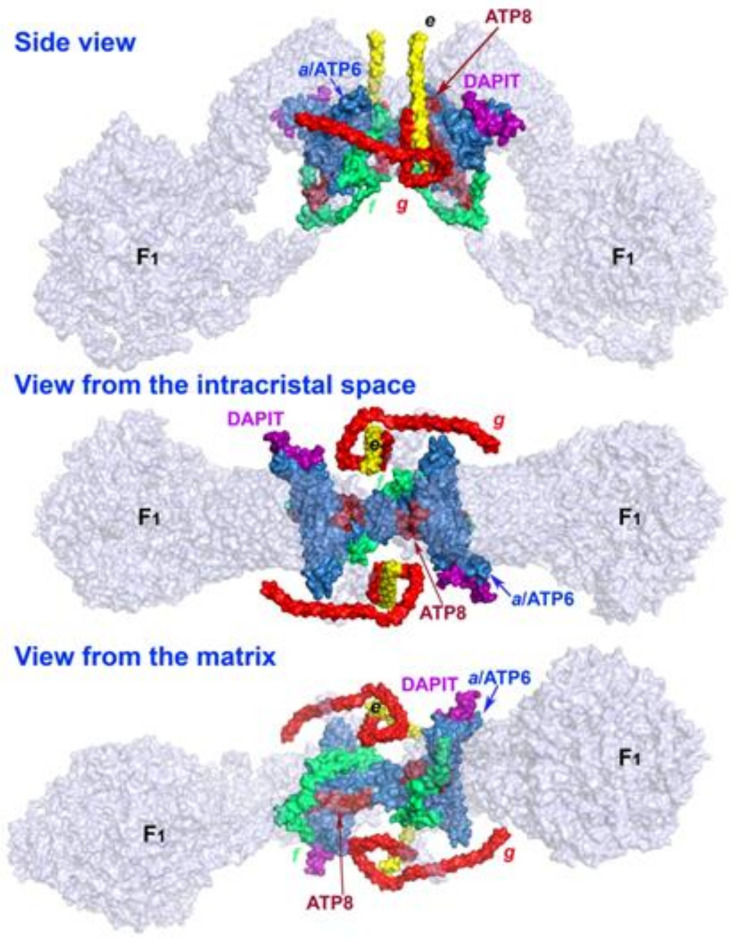
Figure 1.
DAPIT location within the ATP synthase structure. Neighbor subunits of DAPIT are shown within the structure of membrane FO-sector of the ATP synthase. DAPIT and the surrounding subunits are depicted in color, the remaining subunits are transparent. A side view represents a transversal section of the crista rim; a “bottom” view is from the intracristal space directly viewing the inner sharpest IMM bend, when one considers the F1 moiety being positioned at the top. A top view is also the top view for the crista rim and the top of the sharp edge of the IMM bent. The ATP synthase structure was derived from the atomic model for the dimeric FO region of mitochondrial ATP synthase published by Guo H. et al. [37], pdb code 6b8 h. The structure was visualized using the PyMOL Molecular Graphics System, Version 1.8 Schrödinger, LLC.

