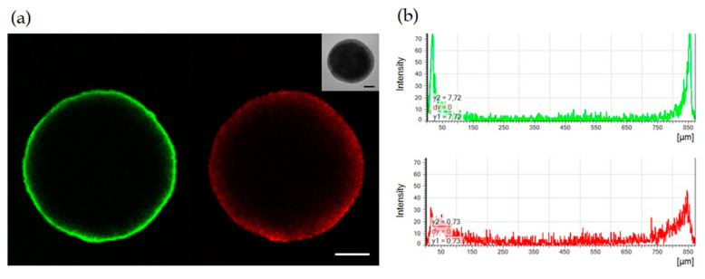
Figure 2.
Confocal microscopy analysis of SCESM spheroid. Seven-day-old SCESM spheroid was stained for the presence of the proliferation marker Ki-67 (anti-Ki-67) and spheroid nuclei were stained with 7-amino-actinomycin D (7-AAD). (a) Confocal microscope fluorescent and visible light images are presented. Green fluorescence represents Ki-67 and red fluorescence represents 7-AAD staining. Scale bars correspond to 200 μm; (b) Histograms of the fluorescence intensity across the SCESM spheroid diameter are presented; the green histogram displays Ki-67 and the red histogram displays the 7-AAD fluorescence intensity across the SCESM spheroid diameter.

