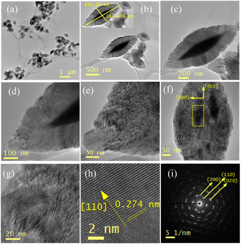
Figure 5.
High resolution transmission electron microscope (HR-TEM) and selected area electron diffraction (SAED) analysis of CuO nanopetals: (a) large scale and well-defined monodispersed CuO nanopetals image on TEM grid. (b,c) TEM image of well-stabilized single CuO nanopetal, showing their length and diameter. (d–g) HR-TEM image of single nanopetal with growth direction exhibiting sharp tips with the clear and smooth surfaces. (h) d-spacing of single CuO nanopetal. (i) SAED analysis taken from the rectangle part in Figure 5.

