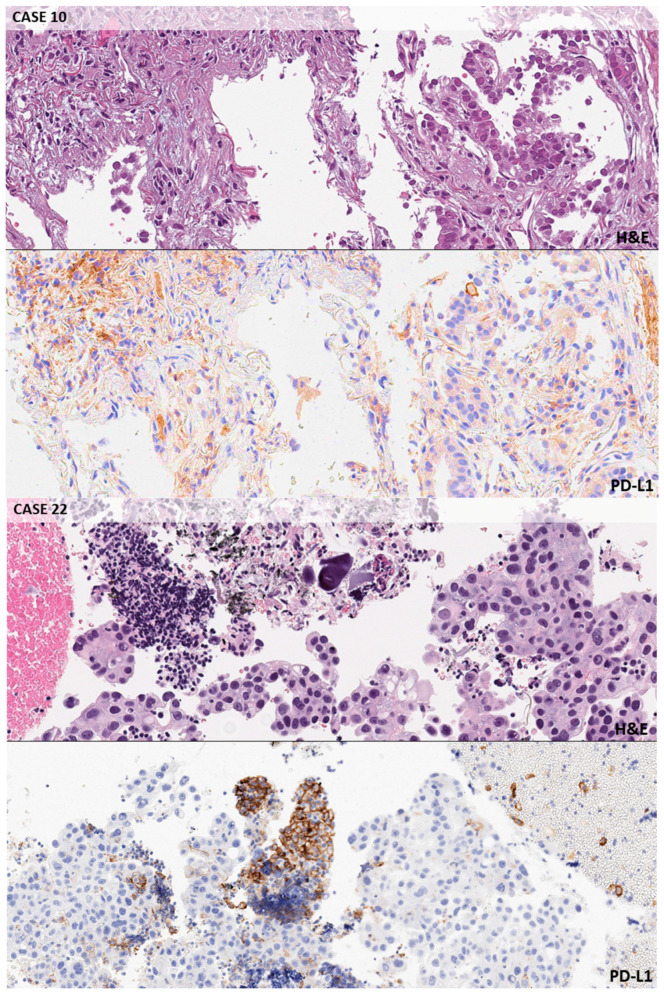
Figure 1.
Negative cases. Exemplificative false positive background in macrophages and inflammatory cells in a lung biopsy. Case n. 10 (original magnification 10×, 22c3): PDL1 TPS < 1%; look at the background staining in macrophages peritumoral cells. CASE n. 22 (10×, 22c3): PDL1 TPS < 1%; the application of a careful magnification rule allows to classify as macrophages this group of PD-L1 strong positive cells.

