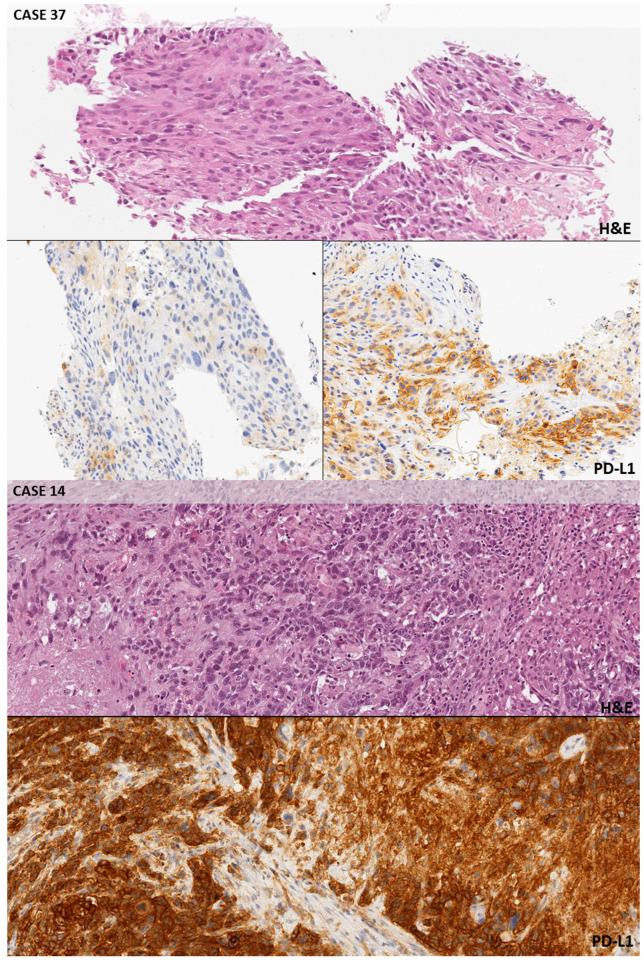
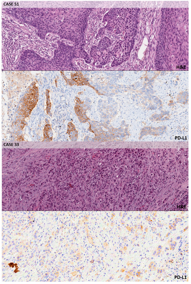
Figure 2.
Intermediate and strong positive cases. Case n. 37 (10×, SP263): PD-L1 TPS > 50%; heterogeneous PD-L1 expression throughout the same tumor, with areas only showing faint background staining in non-neoplastic cells (bottom row, left) along with areas characterized by strong and complete membrane staining in the cancerous elements (bottom row, right). A challenging decision was jointly obtained to decide the exact threshold. Case n. 14 (10×, 22c3): PD-L1 TPS > 50%; all participants correctly ascribed this case to the ‘strong expression’ category due to the diffuse presence of PD-L1 staining in the tumor cells. Case n. 51 (10×, SP263): PD-L1 TPS 1–49%; heterogeneous PD-L1 expression with strong staining cells closely intermixed with faintly staining or negative cells. Case n. 33 (10×, 22C3): PD-L1 TPS 1–49%; a few of the neoplastic elements from this poorly differentiated adenocarcinoma showed a moderate membrane staining with the antibody, so this case was classified as an example of ‘intermediate expression’.


