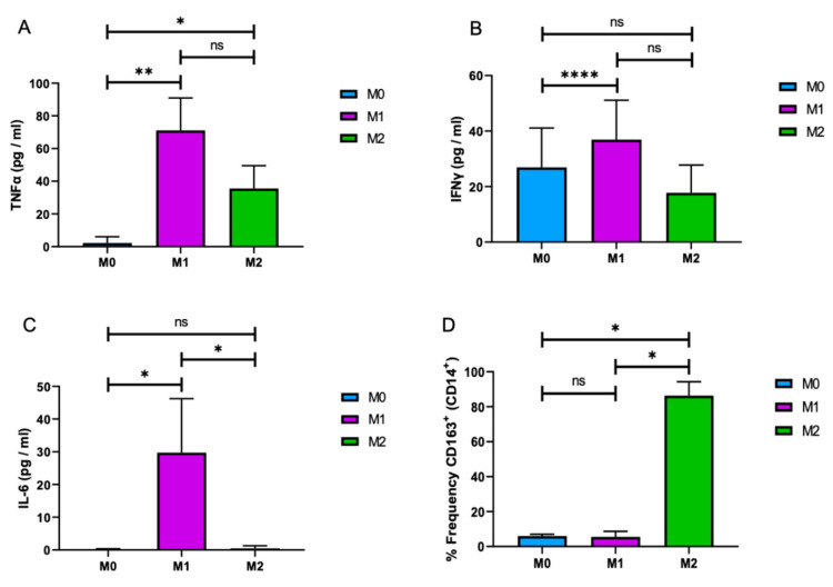
Figure 2.
Differential cytokine expression of M1 and M2 macrophages. M1 and M2 macrophages were differentiated from MonoMac-1 cells (M0), and growth media of the cells was subjected to the Enzyme-linked Immunosorbent Assay (ELISA) for TNF-α (A), IFN-γ (B), and IL-6 (C). (D) M0, M1, and M2 cells were analyzed by flow cytometry for the frequency of CD163+ and CD14+ macrophages. (n = 3; * p ≤ 0.05, ** p ≤ 0.01, and **** p ≤ 0.001).

