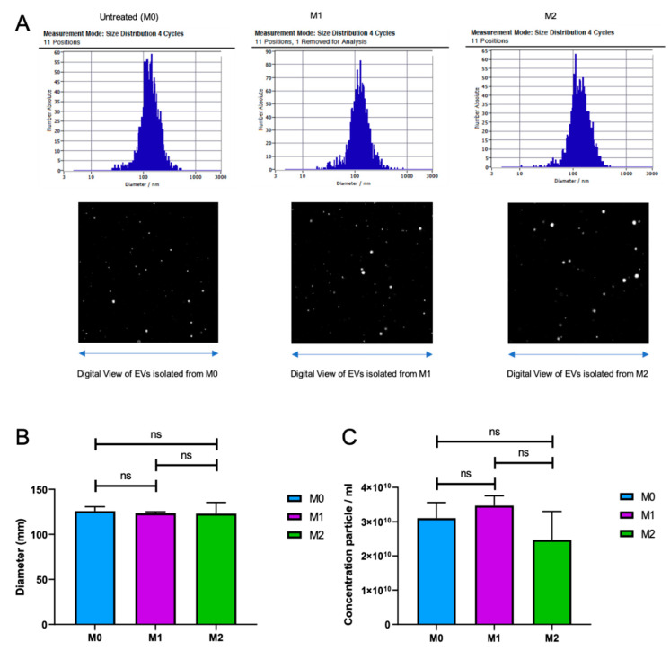
Figure 3.
Extracellular vesicles (EVs) isolated from M0, M1, and M2 cells are indistinguishable. (A) Zeta view analysis of the EVs extracted from M0, M1, and M2 macrophages. Size distribution in 4 cycles—11 positions and digital view of EVs from each cell type are shown. The size (diameter) (B) and concentration (particle number) (C) of EVs extracted from M0, M1, and M2 macrophages are shown as bar graphs. (n = 3).

