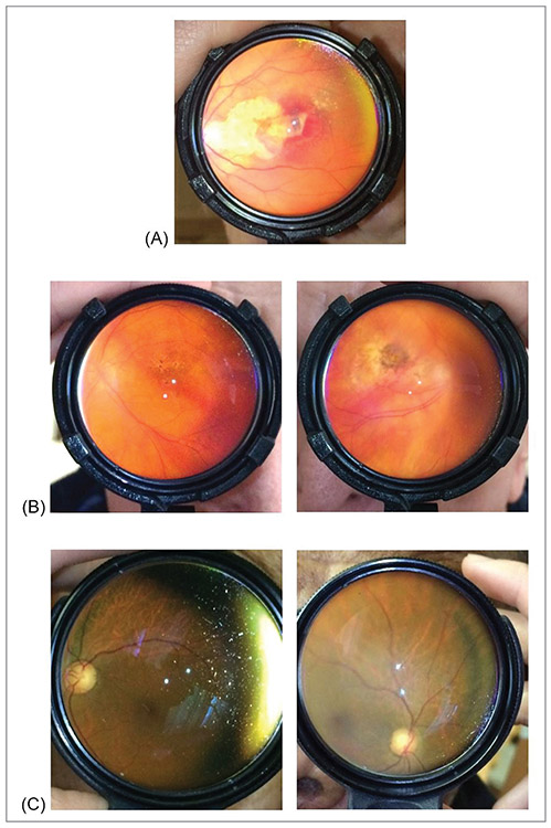
Figure 4.
Example posterior segment fundus photographs taken with Paxos Scope showing (A) neovascular age-related macular degeneration (AMD) with active choroidal neovascular membrane and hemorrhage, (B) high-risk intermediate AMD and advanced dry AMD with geographic atrophy, and (C) asymmetric disc cupping.

