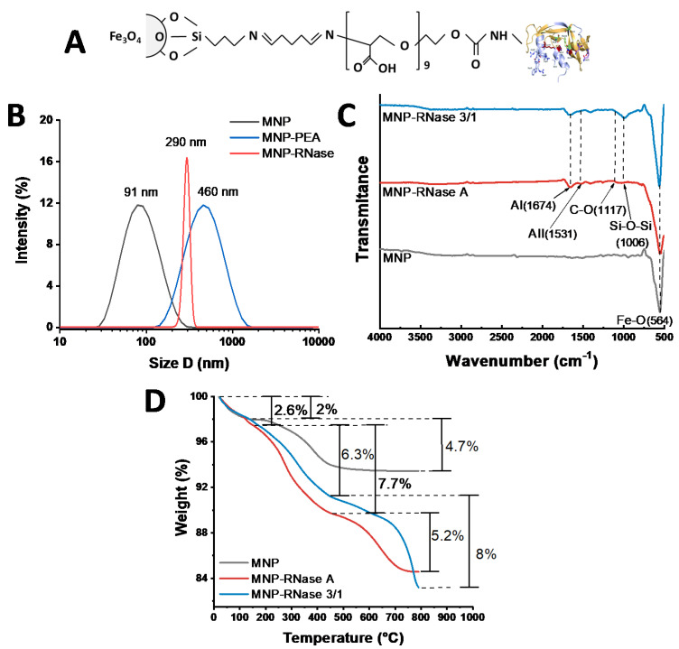
Figure 1.
(A) Schematic representation of the chemical structure of the MNP-RNase bionanoconjugate. (B) DLS histogram for the size distribution of magnetite nanoparticles before (gray), PEA coated NPs (blue) and after immobilization MNP-RNase (red) nanobioconjugates. (C) FTIR spectra of bare magnetite (gray), MNP-RNase A nanobioconjugates (red), and magnetite-RNase 3/1 nanobioconjugates (blue). AI—Amide I and AII—Amide II. (D) TGA thermograms of magnetite (gray) and MNP-RNase A nanobioconjugates (red), and magnetite-RNase 3/1 nanobioconjugates (blue). The first weight loss steps (2.0%) represent the dehydration of the samples. Second weight loss steps (4.7%, 7.7%, and 6.3%) correspond to physically adsorbed organic solvents. The final weight loss steps (5.2% and 8.0%) are attributed to the detachment of RNase A and RNase 3/1 from the surface of magnetite nanoparticles.

