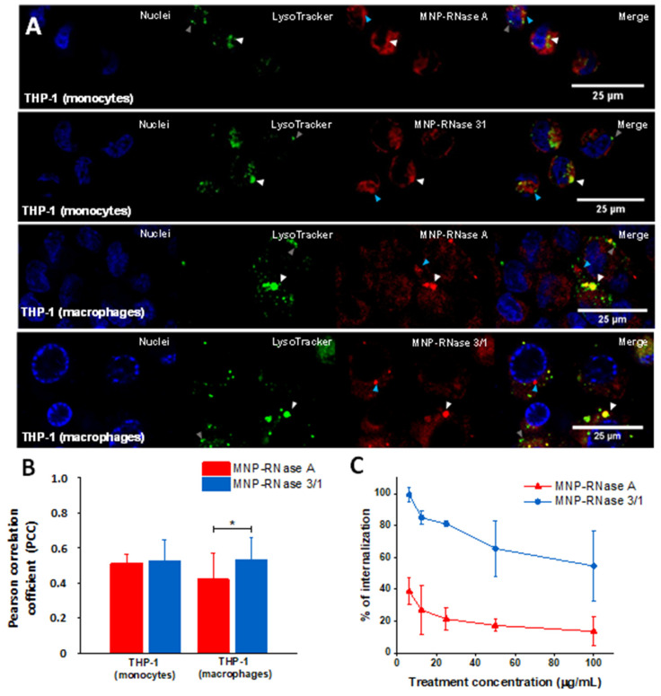
Figure 6.
(A) Confocal microscopy of MNPs-RNase uptake into THP-1 and induced-macrophage cells. Both cell types were stained with Hoechst (blue) to visualize the nuclei. The nanobioconjugates were labeled with Rhodamine B (red), while lysosomes were stained with LysoTracker (green). White triangles indicate lysosomes, the blue ones pointed to bionanoconjugate accumulation outside lysosomal compartments and the gray ones represent empty lysosomal compartments. (B) Colocalization ratio was determined by image processing via the open access software Image J/Fiji® (n = 20 fields with at least 5 cells, * p-value < 0.05). (C) Internalization percentage of MNPs-RNase A and 3/1 nanobioconjugates in THP-1 cells. At low concentrations of nanobioconjugates, uptake by the cells reaches between 40% and 100%. However, increasing the concentration saturates the cells and consequently, the nanobioconjugates uptake reduces to about 10 to 55%.

