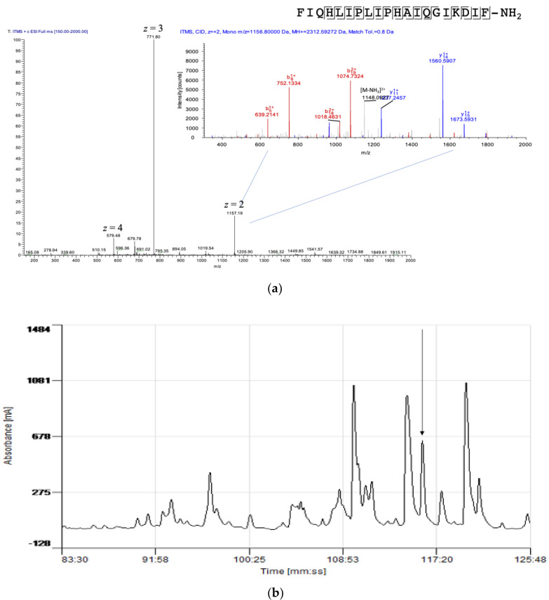
Figure 3.
(a) Electrospray ion-trap MS/MS fragmentation analysis of kassinatuerin-3 in the skin secretion. The full mass scan exhibited multiple positively charged ions of kassinatuerin-3. The doubly charged precursor ion was subjected to MS/MS fragmentation (in the upper right-hand corner). The observed singly and doubly charged b-ion and y-ion fragments m/z ratios are colored as red and blue, respectively. (Note that, in the sequence call, I/L residues are isobaric and cannot be differentiated here. The assignation of I/L is done by reference to the cloned precursor template.) (b) Region of RP–HPLC chromatogram of the skin secretion of K. senegalensis, with an arrow indicating elution/retention time of the fraction (#116) containing the antimicrobial peptide, kassinatuerin-3. The Ɣ-axis indicates milli-absorbance units at λ = 214nm.

