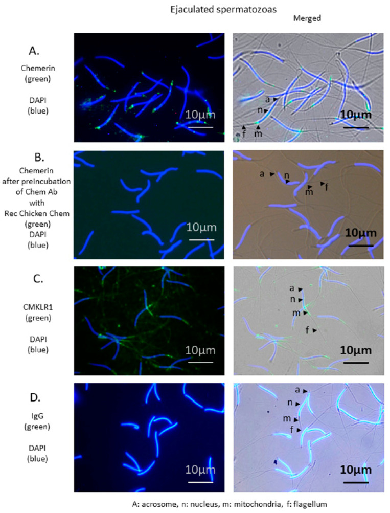
Figure 6.
Localization of chemerin and its receptor, CMKLR1 in spermatozoa of adult roosters (40-week-old). Immunofluorescence on spermatozoa using chemerin (A) and CMKLR1 (C) antibodies shows staining of the mid-piece. Nuclei are stained in blue (DAPI), and specific antigens are stained in green. A: Acrosome, n: Nucleus, m: Mitochondria, f: Flagellum. ejaculated spermatozoa Scale bar = 10 µm. (B) shows the abolition of specific staining following pre-absorption of chicken chemerin antibody with the recombinant chicken chemerin. (D) IgG was used as a negative control. Immunostainings shown are representative of spermatozoa from three different animals.

