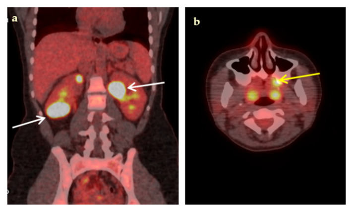
Figure 2.
An 18-year-old woman presented with a 3-week fever, elevated inflammatory blood markers (ESR 77 mm/h, CRP 9.3 mg/dL) and an abdominal CT scan suggestive of possible renal abscesses. Coronal fused 18F-FDG-PET/CT image (a), demonstrated multiple hypodense, highly hypermetabolic lesions in both kidneys, the largest (white arrows) in the lower pole of the right kidney (SUVmax 35.3) and in the upper pole of the left kidney (SUVmax 34.4), compatible with renal abscesses, confirmed by biopsy. Transaxial fused 18F-FDG-PET/CT image (b) demonstrated high 18F-FDG paradental uptake in the left maxilla (yellow arrow) raising concern for hematogenous spread of dental infection. Complementary focused interrogation revealed a history of a painful, undertreated dental condition of the left maxilla preceding fever.

