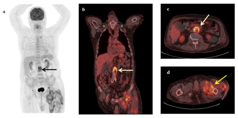
Figure 3.
A 74-year-old man with a medical history of aortobiiliac vascular prosthesis because of an asymptomatic aneurysm 5 months ago, presented with a 2-month fever, increased inflammatory blood markers (ESR 83 mm/h, CRP 19.2 mg/dL) and intramuscular fluid collections in the left thigh, (revealed in a CT for localized pain). Maximum intensity projection 18F-FDG-PET (a), coronal fused (b) and transaxial fused 18F-FDG-PET/CT images at the level of the L3 vertebra (c), demonstrated increased metabolic activity in the wall of the abdominal aneurysm (arrows) at that level (SUVmax 7.0), suspicious of infection. In addition, transaxial fused FDG-PET/CT image at the level of thighs (d) revealed intramuscular hypermetabolic collections with air bubbles in the left thigh, suspicious of abscesses (yellow arrow). Vascular graft infection was confirmed by histopathology after removal of the aortic graft (Klebsiella pneumoniae, Pseudomonas aeruginosa).

