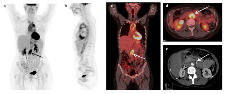
Figure 4.
A 52-year-old woman presented with prolonged fever (over 6 months) and increased inflammatory blood markers (ESR 76 mm/h, CRP 9.3 mg/dL). The patient had a medical history of breast cancer seven years ago. Maximum-intensity projection 18F-FDG-PET (a), sagittal 18F-FDG-PET (b), coronal fused 18F-FDG-PET/CT (c), and transaxial fused FDG-PET/CT images at the level of the renal vessels (d) demonstrated increased metabolic activity within the wall of the thoracic and abdominal aorta, extending to the subclavian arteries, more intense at the root of the aorta and above the aortic bifurcation (arrows). The findings were compatible with vasculitis of large- and medium-sized arteries, mainly affecting the aorta (panaortitis). Transverse section of the CT angiography at the level of renal vessels (e) showed thickening of the aorta wall, more pronounced at the level below the renal vessels (arrow), but with no evidence of narrowing of the aortic lumen. A diagnosis of large vessel vasculitis/Takayasu’s arteritis was made, and fever resolved after treatment with corticosteroids.

