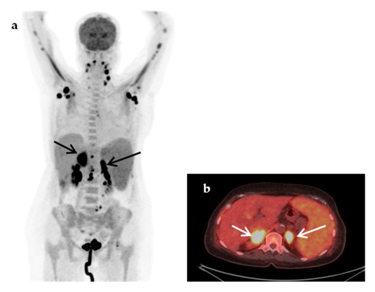
Figure 5.
A 75-year-old woman presented with a 4-week fever associated with malaise and weight loss. The abdominal CT revealed splenomegaly and adrenal masses. Maximum-intensity projection (MIP) 18F-FDG-PET (a) showed extensive highly hypermetabolic lymphadenopathy, cervical, axillary (SUVmax 22.7) and abdominal and hypermetabolic (SUVmax 16.8) adrenal masses (fused transaxial 18F-FDG-PET/CT image (b), arrows), hypermetabolic hepatic lesions (at least two) and multiple hypermetabolic bone metastases and splenomegaly with diffuse homogeneously increased metabolic activity. The findings were suspicious of lymphoma. A diagnosis of non-Hodgkin’s lymphoma was confirmed by histopathology after biopsy of an axillary lymph node.

