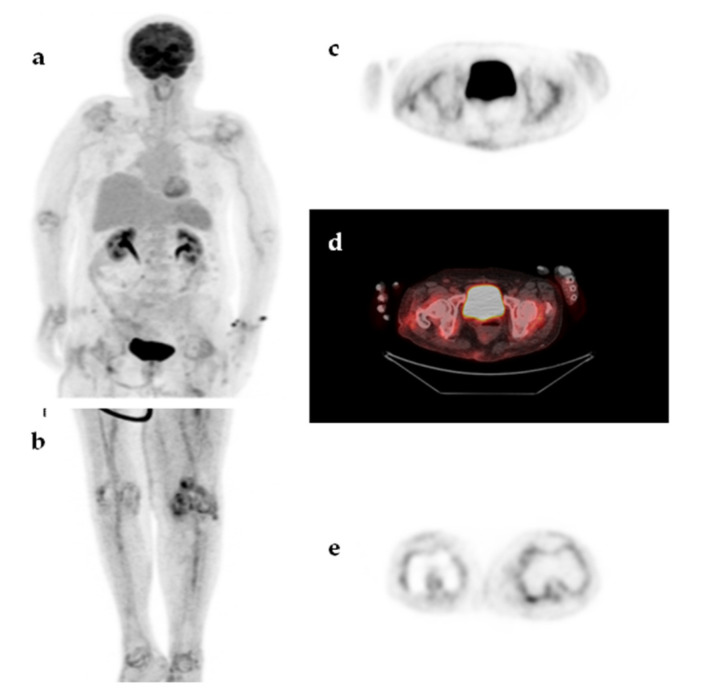
Figure 6.
An 85-year-old woman presented with a 2-month fever. The patient had a history of total left knee arthroplasty with no signs of loosening or infection. MIP 18F-FDG-PET image (a,b), transaxial 18F-FDG-PET and 18F-FDG-PET/CT images at the level of hips (c,d) and transaxial 18F-FDG-PET image at the level of knees (e) demonstrated diffuse, symmetric, moderately increased 18F-FDG uptake, in the large peripheral joints (shoulders, hips, knees) accompanied by increased 18F-FDG uptake along the medium-sized arteries (axillary, humeral, femoral, and tibial arteries). The findings were compatible with polymyalgia rheumatica and the fever resolved upon treatment with corticosteroids.

