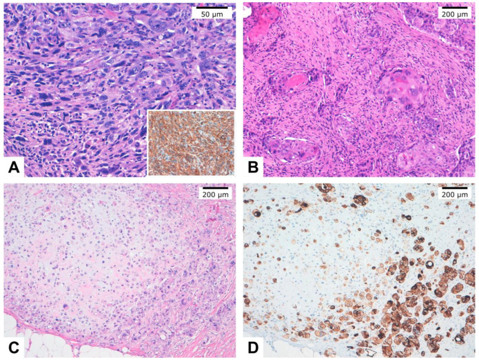
Figure 1.
Histopathological images of different metaplastic breast carcinoma subtypes. (A). Spindle cell carcinoma entirely composed of neoplastic spindle cells. Inset: Immunohistochemistry showed cytokeratin AE1/AE3 diffuse and intense staining in tumor cells. (B). Squamous cell carcinoma with keratin pearls. (C). Metaplastic carcinoma with mesenchymal (chondroid) differentiation (matrix-producing carcinoma). (D). Immunohistochemistry of case C showed cytokeratin AE1/AE3 expression staining in tumor cells.

