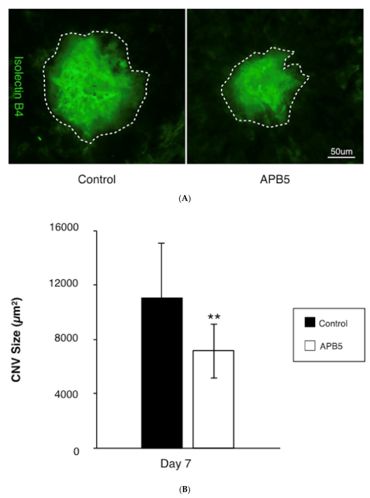
Figure 2.
Suppression of CNV formation by PDGFR-β blockade (A) Representative micrographs of CNV lesions (isolectin B4, green) in the RPE-choroid flat mounts at post-laser day 7 from mice treated with IgG control or APB5 antibody, respectively. The white dotted line indicates the outline of CNV lesion. Scale bar: 50 μm. (B) Quantification analysis of the size of CNV. (** p < 0.01, n = 12 eyes).

