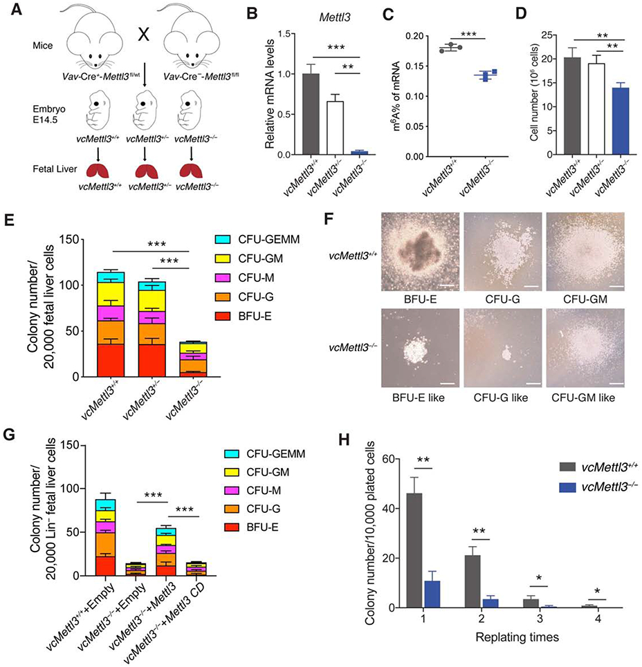
Figure 1. Loss of METTL3 results in dysfunctional fetal liver hematopoietic stem cells.

(A) Breeding scheme to obtain Vav-Cre-Mettl3fl/fl mice vcMettl3+/+, vcMettl3+/−, and vcMettl3−/− E14.5 fetal livers.
(B) Quantitation of Mettl3 expression via Q-RT-PCR, normalized to Gapdh (n=3 biological replicates per group).
(C) Quantitation of m6A of mRNA in vcMettl3+/+ and vcMettl3−/− E14.5 fetal livers via ELISA (n=3 biological replicates per group).
(D) Fetal liver cell numbers at E14.5 in vcMettl3+/+, vcMettl3+/− and vcMettl3−/− embryos (n=5 biological replicates per group), counted after mechanical dissociation and red blood cell lysis.
(E, F) Assessment of colony forming unit (CFU) potential of vcMettl3+/+, vcMettl3+/− and vcMettl3−/− E14.5 fetal liver cells (E) (n=3 biological replicates per group). Demonstration of typical colonies in vcMettl3+/+ and vcMettl3−/− cultures (F). CFU-GEMM, CFU-granulocyte, erythrocyte, monocyte, megakaryocyte; CFU-GM, CFU-granulocyte, macrophage; CFU-M, CFU-macrophage; CFU-G, CFU-granulocyte; BFU-E, burst-forming unit-erythroid. Scale bar, 100 μm.
(G) CFUs of E14.5 lineage depleted fetal liver cells transduced with empty, Mettl3 and catalytically dead Mettl3 (Mettl3 CD) expressing retroviral vectors (n=3 biological replicates per group).
(H) Serial colony replating assay enumerating colonies/10,000 plated cells (n=3 biological replicates per group).
Data are represented as mean ± SEM, and representative of at least two independent experiments; The P values were calculated using two-tailed Student's t test. * p<0.05, ** p<0.01, *** p<0.001.
See also Figure S1.
