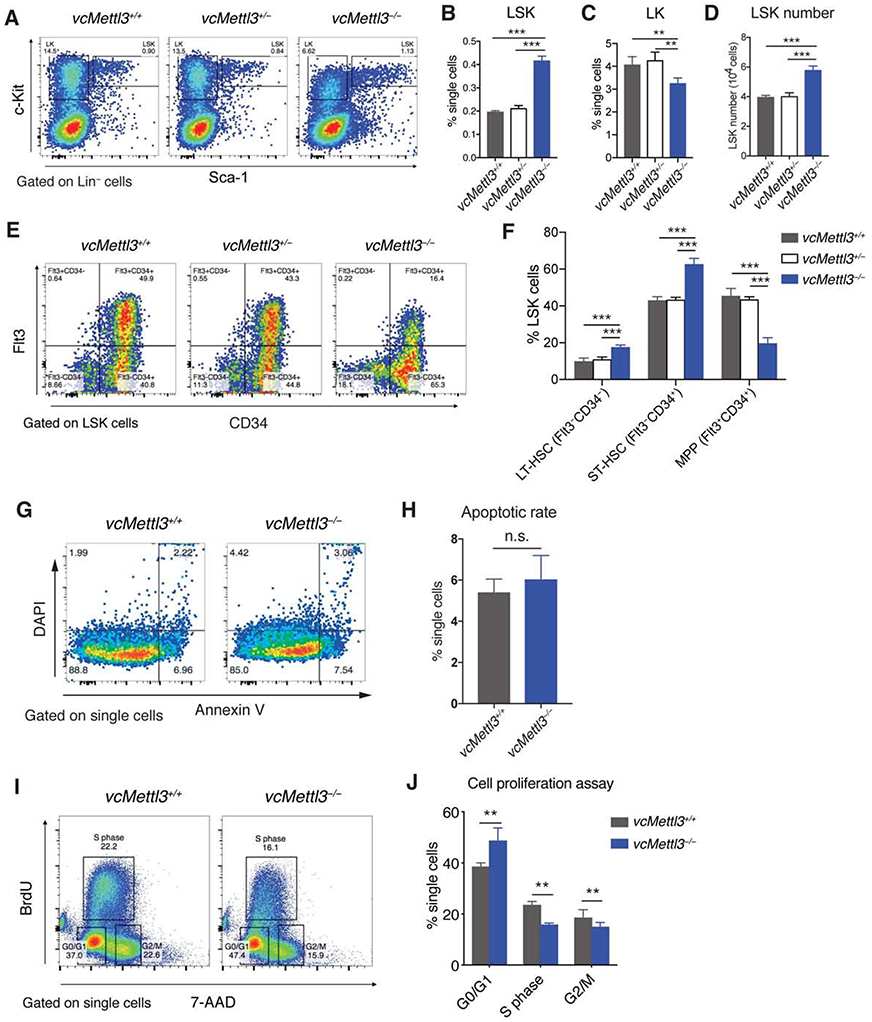
Figure 2. Mettl3-deficient fetal liver hematopoietic stem and progenitor cells show aberrant differentiation trajectory and impaired proliferation.

(A-D) Determination of LSK and LK cell distribution in vcMettl3+/+, vcMettl3+/− and vcMettl3−/− E14.5 fetal livers by flow cytometry (A). Quantitation relative to single cell population of LSK cells (B) and LK cells (C), and absolute LSK cell numbers per fetal liver (D; n=5 biological replicates per group).
(E and F) Determination (E) and quantification (F) of phenotypic hematopoietic stem cell subsets in vcMettl3+/+ and vcMettl3−/− E14.5 fetal livers via flow-cytometric identification of long-term (LT) (Flt3−CD34− LSK) and short-term (ST) (Flt3−CD34+ LSK) hematopoietic stem cells (HSCs) and multipotent progenitors (MPPs) (Flt3+CD34+ LSK; n = 3 biological replicates per group).
(G and H) Determination (G) and quantification (H) of apoptotic rate in fetal livers via annexin V staining (n = 3 biological replicates per group).
(I and J) Assessment (I) and quantification (J) of fetal liver cell proliferation via BrdU uptake and 7AAD staining of DNA content (n = 3 biological replicates per group).
Data are represented as mean ± SEM and representative of at least three independent experiments; the p values in (B)–(D) and (H) were calculated using two-tailed Student’s t test. n.s., not statistically significant; **p < 0.01; ***p < 0.001. The p values in (F) and (J) were calculated using two-way ANOVA. **p < 0.01; ***p < 0.001.
See also Figures S2 and S3.
