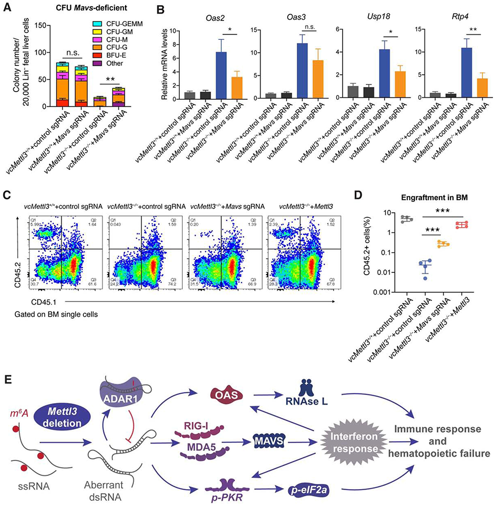
Figure 7. Inhibition of the innate immune response partially rescues hematopoietic failure secondary to Mettl3 deletion.

(A) Determination of the effect of CRISPR/Cas9 mediated Mavs deletion on colony formation in vcMettl3−/− compared to vcMettl3+/+ Lin− fetal liver cells (n=3 biological replicates per group).
(B) qRT-PCR measurement of Oas and interferon response gene expression secondary to control versus targeted sgRNA-mediated deletion of Mavs in vcMettl3−/− compared to vcMettl3+/+ fetal liver cells, normalized to Gapdh (n = 3 biological replicates per group).
(C and D) Determination (C) and quantification (D) of the engraftment of vcMettl3−/− cells with CRISPR-Cas9-mediated deletion of Mavs or overexpression of Mettl3 6 weeks post-transplantation via flow cytometric detection of CD45.2 fetal liver cells versus CD45.1 control cells (n = 4 separate mice per group). One independent experiment is shown.
(E) Model of the Mettl3 deletion-induced dsRNA-mediated innate immune response.
Data are represented as mean ± SEM, and representative of at least two independent experiments unless stated otherwise; The P values were calculated using two-tailed Student's t test. n.s. not statistically significant, * p<0.05, ** p<0.01, *** p<0.001.
See also Figure S7.
