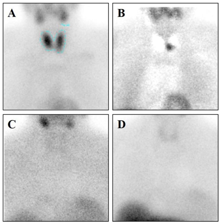
Figure 1.
[99mTc]Sestamibi Single Photon Emission Computed Tomography (SPECT) Analysis. (A) Image shows [99mTc]Sestamibi uptake in a 54-year-old woman with primary hyperparathyroidism. A parathyroid carcinoma (0.6 cm) was identified after the surgery by histological analysis. (B) To evaluate the parathyroid sestamibi uptake, that of the thyroid has been subtracted (C) Image displays no [99mTc]Sestamibi uptake in a 68-year-old woman with primary hyperparathyroidism. A parathyroid hyperplasia (0.2 cm) was identified after the surgery by histological analysis. (D) To evaluate the parathyroid sestamibi uptake, that of the thyroid has been subtracted.

