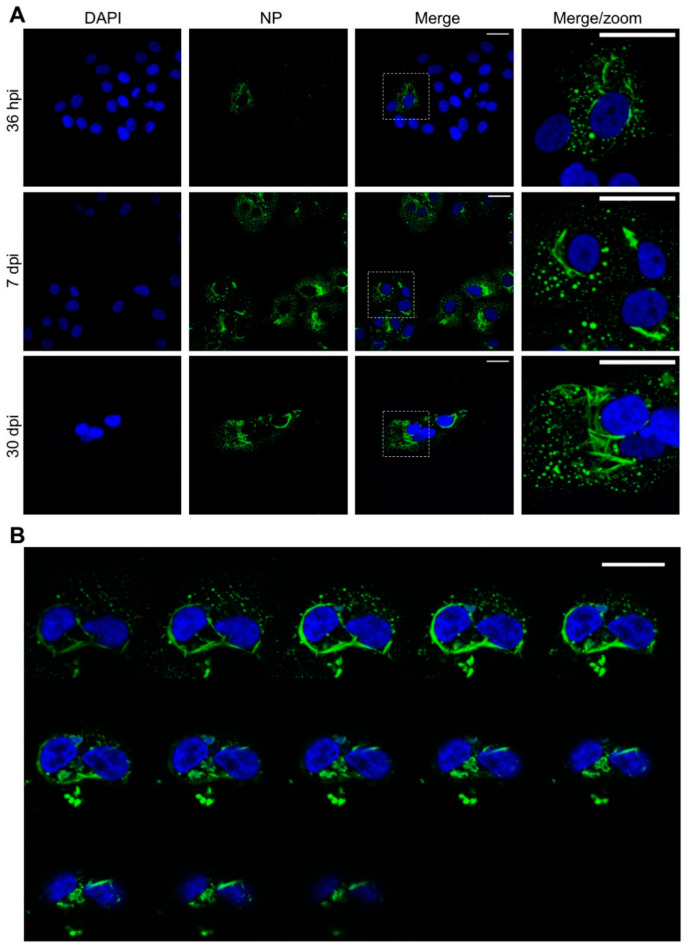
Figure 1.
Confocal images showing the characteristic morphology of Tula virus (TULV) nucleocapsid protein (NP) -stained structures at early, peak and persistent stages of TULV infection in Vero E6 cells. (A) Vero E6 cells were infected with TULV at a multiplicity of infection of 0.1, and the images were taken at 36 h post infection (hpi), 7 days post infection (dpi) and 30 dpi using 40× magnification. Nuclei were stained with DAPI (blue) and TULV NP, detected using NP antisera (green), revealing the characteristic NP punctate and filamentous/tubular structures in the perinuclear region. (B) Successive Z sections of TULV-infected cells at 30 dpi show NP filamentous/tubular structures extending around the nucleus. The scale bars represent 30 μm.

