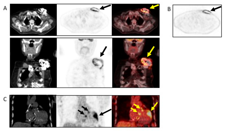
Figure 8.
Example of the use of (18F)FDG PET/CT in a patient with non-Hodgkin’s lymphoma and sudden onset of fever and a positive blood culture for Streptococcus dysgalactiae. The patient underwent TTE and TEE, which were negative. Antimicrobial treatment was started. Due to the lack of clinical response, the patient underwent (18F)FDG PET/CT, which revealed infection, as indicated by the black and yellow arrows, involving the pocket of the device ((A), from left to right, CT, PET, and superimposed PET/CT transaxial (upper panel) and coronal (lower panel and (B) non-attenuated corrected transaxial images) as well as the intracardiac portion of the lead extending to the tricuspid valve ((C), from left to right, noncontrast CT, PET, and fused PET/CT coronal images). Based to the PET/CT findings, the device was extracted and replaced.

