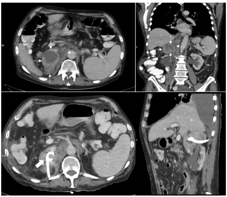
Figure 1.
Upper row: 11 × 6 cm retroperitoneal mass in the right adrenal loge (thin white arrows) with contact to diaphragm, liver and retroperitoneal vessels. The pararenal space is not involved. The mass shows an inhomogeneous enhancement. The right adrenal gland itself cannot be distinguished. Lower row: Mass after puncture and CT guided drainage with a pigtail catheter (thick white arrow).

