Figure 1.

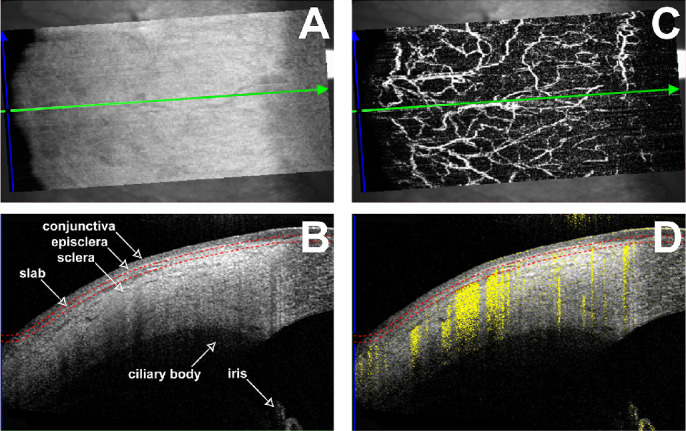
Assessment of conjunctival vessels using anterior OCTA. (A) En face OCT of the temporal quadrant of a right eye corresponding to the conjunctival slab in B. Green and blue arrows refer to the position of the OCT scan. (B) OCT image in the position of the green arrow in A. The two red dotted lines show the depth of the OCTA slab (100 µm depth, 50 µm thickness). (C) En face OCTA revealing the vascular meshwork of the conjunctival slab. (D) OCTA scan in the position of the green arrow in A. The two red dotted lines show the depth of the OCTA slab (100 µm depth, 50 µm thickness). The reflectivity of vessels is shown in yellow within the OCT scan.
