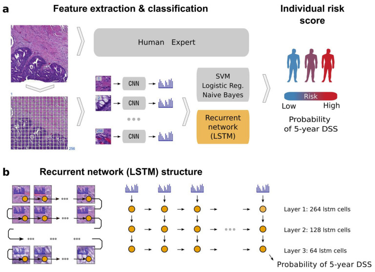
Figure 7.
Example of patient prognosis of CRC patients. Overview of the image analysis pipeline and long short-term memory (LSTM) prognostic model. Images of tissue microarray (TMA) spots are characterized by a pre-trained convolutional neural network (VGG-16) in a tile-wise manner. The VGG-16 network produces a high-dimensional feature vector for each tile from an input image. These features then serve as inputs for classifiers trained to predict five-year disease-specific survival (DSS) (a). The long short-term memory (LSTM) network slides through the entire image of the tissue microarray spot to jointly summarize observed image tiles and predict the patient risk score (b). Figure 1 in Bychkov et al. [49], copyright 2018 the author(s), reprinted from the Scientific Reports published by Nature. This is an open access articles under the term of the Creative Commons attribution-Non Commercial-NoDerivs License.

