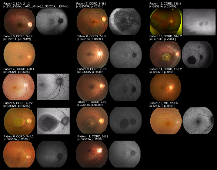
Figure 2.
Fundus photographs and fundus autofluorescence images of 14 patients with GUCY2D-associated retinal disorder (GUCY2D-RD; patients 2–15). Fundus photographs and fundus autofluorescence (FAF) images of the right eyes demonstrated macular atrophy in seven affected subjects (patients 4, 5, 7, 9, 10, 12, 13) with intrachoroidal cavitation in three subjects (patients 5, left; 10, left; 13) and slight fine dots at the macula in two subjects (patients 4, 9). Atrophic change at the posterior pole extending to the periphery was observed in patient 7 and subtle diffuse disturbance at the posterior pole with vessel attenuation was found in patient 7. Normal fundus appearance was noted in five subjects (patients 1, 2, 6, 8, 14). Patient 11 had essentially normal retinal appearance except for optic disk cupping. The atrophic changes were more evident on FAF images. A loss of AF signal at the macula was identified in five subjects (patients 5, 7, 10, 12, 13). Increased AF signal at the macula was observed in five subjects (patients 2, 4, 6, 8, 11). A patchy area of decreased AF signal at the posterior pole extending to the periphery surrounded by a ring of increased AF signal was found in patient 7.

