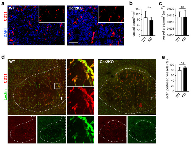
Figure 5.
Glioma vasculature is unaffected by Ccr2-deficiency. (a) Representative images of brain tumor sections of WT and Ccr2KO mice stained for CD31 are depicted (d21). Scale bars 100 µm. (b,c) Graphs indicate vessel count (b) and vascularized area (c) within the tumor tissues (n = 6). (d) Confocal microscopy of brain tumor sections from mice perfused with FITC-Lectin and stained for CD31 (overview, mosaic). Square indicates the magnified region (63×). T, tumor tissue; dashed line, tumor border. (e) The graph presents the percentage of perfused vessels (n = 3–4). ns, not significant (unpaired Student’s t-test).

